The diagnosis of atopic dermatitis is clinical; no reliable markers exist for diagnosis or assessing disease severity.
Learn more about IgE Levels in Atopic Dermatitis here.
Signs and Symptoms
- The chief symptom of atopic dermatitis is pruritus.
- Xerosis is often present.
- Scratching leads to excoriated and lichenified lesions.
- The typical distribution of lesions varies by age (Figures 1–3).
- The appearance of the lesions varies by ethnicity.
- Lightly pigmented skin lesions may appear erythematous or as scaly and/or crusted papules, patches, or thin plaques.
- Erythema is less obvious in the skin of children with more pigmentation. The lesion is more papular and postinflammatory hyper- or hypopigmentation is often present.
Typical Distribution of Lesions
Infants
Infants often have scaly, crusted patches on the cheeks, scalp, trunk, thighs, shins, and outer (extensor) surfaces of the upper and lower extremities (see Figure 1).
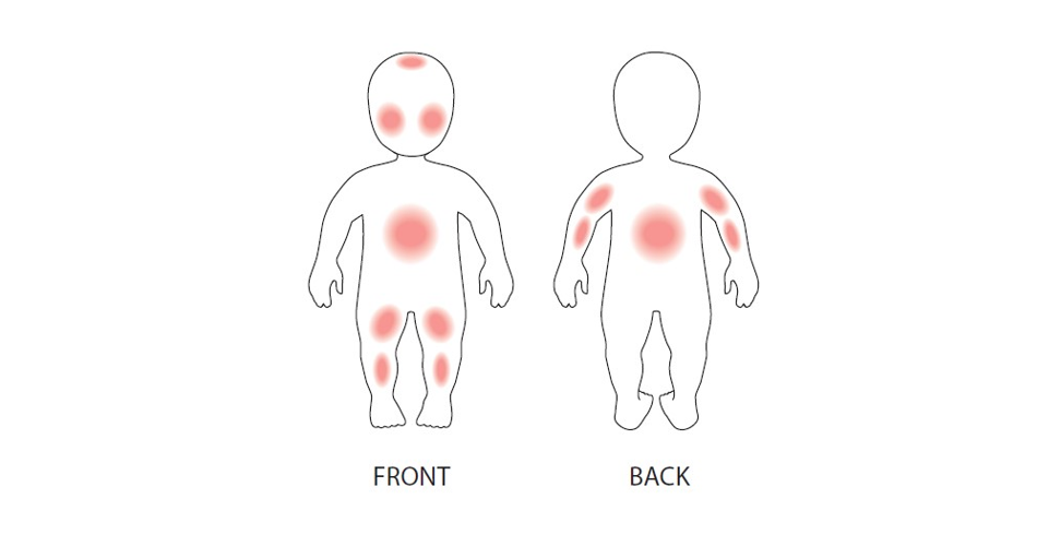
Figure 1: Distribution of skin lesions in infants with atopic dermatitis.
Young Children
Toddlers and young children commonly present with lesions on the scalp, flexural joints (antecubital and popliteal fossae), and the back of the neck (see Figure 2). Some children exhibit round, crusted (nummular) lesions.
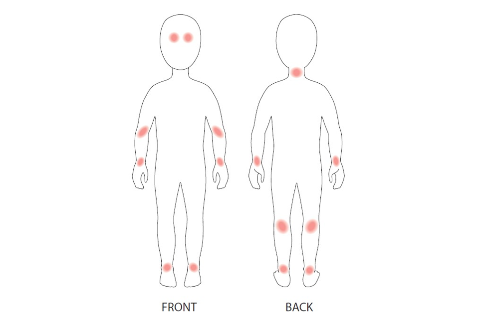
Figure 2: Distribution of skin lesions in young children with atopic dermatitis.
Adolescents and Adults
The distribution of atopic dermatitis lesions in adolescents is flexural but also may include the feet, hands, neck, and face, especially the periorbital area1 (see Figure 3).
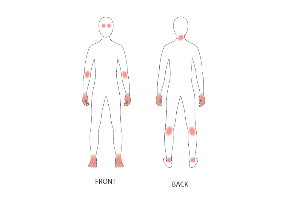
Figure 3: Distribution of skin lesions in adolescents and adults with atopic dermatitis.
Supporting the Diagnosis
Skin conditions often associated with atopic dermatitis that support the diagnosis include
- Morgan folds (atopic pleats): prominent skinfolds located beneath the lower eyelids
- Dry skin (xerosis)
- Hyperlinear palms and soles
- Lichenification: thickened skin with prominent creases
- Keratosis pilaris: papules centered about follicles that have a central core of keratin debris and, at times, surrounding erythema; lesions usually located on the upper outer arms, face, and thighs
- Pityriasis alba: small, poorly defined areas of hypopigmentation located on the face or elsewhere
- Ichthyosis vulgaris: polygonal scales, most commonly involving the lower extremities
Look-alikes
![]()
The development of these resources was made possible with support from Sanofi and Regeneron. The AAP maintains full independence in its editorial and strategic activities. Financial supporters have no influence over AAP content, policies, or leadership decisions.
Last Updated
06/11/2021
Source
American Academy of Pediatrics